Shoulder Resources: Dr. Rudzki’s great love and focus within sports medicine is the shoulder. This is demonstrated by his research, teaching, publications and presentations on partial rotator cuff tears, rotator cuff vascular supply and it’s role in rotator cuff disease, clavicle fractures, and suture anchors. Dr. Rudzki has the honor and privilege of being a member of the American Shoulder & Elbow Surgeons. This highly selective academic society is “made up of leading national and international Orthopaedic surgeons who specialize in surgery of the shoulder and elbow.”
Several case studies, patient testimonials, and blog posts can be found through this portion of our site to help educate patients about common shoulder problems, treatment options, and rehabilitation considerations. Additional information may be gained from visiting links to our YouTube Site, Lectures & Presentations, Research & Publications, and External Sources.
Ruth, a 55 year-old female professional performance pianist and instructor, injured her shoulder in June of 2017 when she fell forward onto outstretched hands while running. The fall resulted in a three-part, displaced proximal humerus fracture to her left shoulder. The x-ray (left) shows the displaced bone fragments of the humeral head.
Work up:
In addition to the initial x-ray, a CT scan with digital 3D reconstruction was ordered to examine the bone quality and see the fracture fragments in more accurate detail. The 3D portion of the CT scan can be seen below. While in some cases, proximal humerus fractures are able to heal with conservative management if the bones are close enough to proper alignment, open reduction internal fixation (ORIF) surgery can ensure that the bones are held in the right place for healing. Given the nature of Ruth’s pianist career, she opted to have the ORIF surgery to increase her likelihood of proper alignment for healing and her chances of a full recovery.
Surgery:
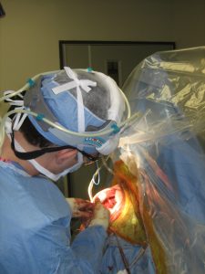
Ruth underwent an open reduction internal fixation (ORIF) of her proximal humerus fracture the next week at Sibley Memorial Hospital. A Synthes locking plate was used to secure the bone fragments in proper anatomic alignment for optimal healing. The surgery generally takes 2.5 hours and can be done as an outpatient procedure. The x-ray on the right is from Ruth’s initial post-operative visit two days after surgery, and you can see that her staples have not yet been removed. Typically, the staples are removed two weeks after surgery.
Recovery:
Ruth started physical therapy approximately 4 weeks after the procedure. She was in a sling for six weeks following the operation. Physical therapy focused on gentle motion restoration and progressed to strengthening. Ruth is now almost one year out from surgery, and she is back to playing the piano. Her range of motion is recovered. As you can see from the pictures below, she is quite happy about her progress.
Related Posts
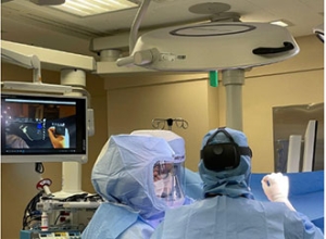
New Technology in Shoulder Replacement: Augmented Reality
Posted on: 21-Sep-2021
Augmented reality is making a place in medicine. Dr. Jonas Rudzki, orthopaedic shoulder specialist, is the first surgeon in DC, Maryland, and Virginia to use a state of the art augmented reality program from Stryker/Tornier
A Triumphant Return to Golf After Shoulder Replacement
Posted on: 25-Oct-2019
J.D. came into the office with shoulder arthritis treated conservatively for years. As an avid golfer and basketball player, he was inhibited from his activities by arthritis. A CT scan was done with 3D images
Triathlete Returns to Competition after Rotator Cuff Revision Repair
Posted on: 10-Mar-2019
“I am supposed to do an Ironman with the oldest grandson when he turns 18…I will be 72) Thank you goes to Dr. Rudzki for doing such an amazing surgical repair on my shoulder