Dr. Rudzki had the privilege of training in Sports Medicine and developing his knee surgical skills at Washington University in St. Louis and the Hospital for Special Surgery in New York City. He has performed high-caliber research on cartilage transplantation as well as ACL injury in NHL players and written articles on bioabsorbable knee implants and fracture fixation about the knee.
Dr. Rudzki has a high level of expertise in the performance of arthroscopic ligament reconstruction, cartilage restoration, complex meniscal repair, treatment of patellar instability, osteotomy and fracture treatment about the knee in a wide range of athletes from adolescents to professionals. He serves on the clinical faculty of the George Washington University School of Medicine teaching medical students and orthopaedic surgical residents. As a consultant for Arthrex and Stryker, Dr. Rudzki engages heavily in medical education to train surgeons and product specialists on emerging techniques and engages with engineers on product design teams to help develop better tools and techniques. As a member of the AAOS Evaluation Committee, he spent several years writing questions on knee injuries and surgical treatments for surgeons in practice and in training.
Posted in: Case Studies, Featured, Knee Resources, Patient Testimonials, Reviews | Tags: Arthroscopy, Discoid Meniscus, Ice Hockey, Knee, Lacrosse, Return to Play, surgery | Posted on: 14-Jun-2017 Chief Complaint/Injury
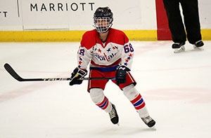
9 year-old male hockey player presents for evaluation of lateral knee pain with intermittent swelling and mechanical symptoms.
Workup/Images
MRI Reveals a Discoid Lateral Meniscus with a central tear. The MRI appearance of a discoid lateral meniscus is consistent and diagnostic when sequential cuts reveal a homogenous, intact communication between the posterior and anterior horns of the lateral meniscus.
Surgery
The patient undergoes successful arthroscopic partial lateral meniscectomy in a procedure which converts the torn discoid lateral meniscus into a normal functional lateral meniscus. This procedure is performed through two small incisions which measure less than 1 cm and enable placement of a high-definition fiber optic video camera. Patients experience mild, transient pain and go home within 1-2 hours after surgery.
Result
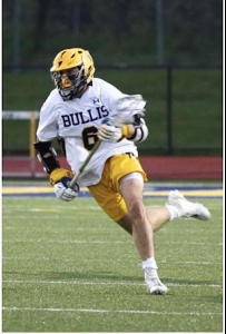
The patient participates in a comprehensive rehabilitation protocol and makes a complete recovery with a full return to sports including travel hockey and lacrosse. Approximately 5 years later, he has competed at the highest levels of Tier 1 elite youth hockey in the U.S. and Canada. This success is a result of his dedication to physical therapy and the collaboration between our patient, his parents, physical therapists, coaches and training specialists.
Discoid Lateral Meniscus is a developmental variant in which a normal semi-circular meniscus fails to form. These may be a symptomatic however they commonly will result in symptoms of swelling, catching, and locking. Surgery typically involves an arthroscopic saucerization or resection of the central portion of the meniscal tissue. It may also involve placement of sutures to secure or stabilize the remaining meniscal tissue. Careful evaluation with a thorough history, physical exam and appropriate diagnostic imaging is essential to help develop an optimal treatment plan.